Broken Sleep and Next-Morning Brain Scans
A 2026 study gave 37 older adults a home sleep test and a brain PET scan the next morning. More time awake in the night went with lower glucose use in several cortical areas. It is a small study of correlations.
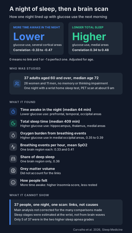
Sleep and brain research often looks years ahead, at memory decline or dementia. A 2026 study from the Mayo Clinic looked at the very next morning.
37 older adults wore a home sleep test for one night and had a brain PET scan a few hours after waking. More time awake in the night went with lower glucose use in several parts of the cortex.
Who was studied?
- 37 adults aged 60 and over from the Mayo Clinic Study of Aging in Minnesota.
- Median age 72, and 26 of them were women.
- None had memory or thinking impairment on a full assessment.
- None were on therapy for sleep apnea.
- On the home test, 16 had no sleep apnea, 16 were in the lowest grade and 5 were in the two higher grades.
What was measured?
- The night. A WatchPAT One, a wrist-worn home sleep test that uses the pulse signal, oximetry and movement. It estimates sleep stages, time awake, breathing events and oxygen levels.
- The morning. An FDG-PET scan, which shows how much glucose each part of the brain is using. The median start time was 9:05 am, about 3.4 hours after waking.
- The researchers compared each sleep measure with glucose use in 47 brain regions, adjusting for age.
A typical night in this group: 409 minutes of sleep, 44 minutes awake after first falling asleep, and a mean SpO2 of 94%.
What did they find?
The results are rank correlations, where 0 means no link and 1 or -1 means a perfect one. The ranges below are from the paper's results section. Its abstract prints slightly narrower ones for time awake (-0.34 to -0.47) and total sleep time (0.39 to 0.42).
- Time awake in the night. More of it went with lower glucose use across the cortex, mainly in prefrontal, lateral temporal and occipital areas (-0.33 to -0.47).
- Total sleep time. Longer sleep went with higher glucose use in medial structures, including the posterior cingulate, hippocampus, thalamus and caudate (0.34 to 0.48).
- Sleep efficiency. A mixed pattern covering areas from both of the above.
- Deep sleep. Linked to one region only (0.36), and that link was no longer statistically significant once grey matter volume was accounted for.
- How people felt. More time awake went with a higher insomnia questionnaire score (0.43). People who felt rested in the morning had a median of 37 minutes awake, against 52 minutes for the rest.
The main links held after adjusting for high blood pressure. They were largely consistent in men and women, though several were no longer statistically significant in the smaller split groups. Grey matter volume did not explain them. The authors say that points to a change in function and not in structure, which might be reversible. They did not test that.
For what broken sleep did to test results the next day in another group, see broken sleep and next-day thinking speed.
What about overnight oxygen?
Two of the three results went the opposite way to what the authors expected.
- Higher oxygen burden went with higher, not lower, glucose use in medial occipital areas (0.35 to 0.39).
- More breathing events per hour (counted with the 4% rule) also went with higher glucose use, in one region (0.33).
- Higher mean SpO2 went with higher glucose use in one region (0.41), which is the direction they expected.
Oxygen burden adds up the depth and length of the oxygen dips that follow breathing events. Here's how it differs from a simple event count.
The authors suggest that blood flow changes after breathing events could carry more tracer to the back of the brain. That is a hypothesis, and the study did not measure blood flow.
What can't it show?
- Cause or direction. It was a cross-sectional study: one night and one scan per person. It can't say whether broken sleep lowers glucose use or something else drives both.
- Certainty. It was an exploratory study of 37 people, and the main analysis did not correct for the many comparisons it made.
- Sleep stages. The wrist device estimates them without recording brain waves, and the authors say it is less accurate at this in older adults. That may be why deep sleep showed so little.
- Drowsiness in the scanner. People were told to stay awake, but the authors can't rule out that some drifted off.
- Sleep apnea. Only 5 of the 37 were above the lowest grade, so the oxygen results rest on a group with few frequent breathing events.
What can you do with this?
Nothing in it changes what a single night of your own data means. The correlations ran from about 0.3 to 0.5 in size, and nobody in the study was followed over time.
If you track your nights, time awake is worth looking at alongside your oxygen numbers. One night varies a lot, so record several before you compare.
If you often wake unrested, tell your doctor.
Not a medical device. Does not diagnose, treat or prevent any condition. Discuss results and changes with a clinician.
References
- Carvalho DZ, Adolf J, Kremen V, McCarter SJ, St Louis EK, Przybelski SA, Liu K, Jones DT, Petersen RC, Jack CR Jr, Lowe VJ, Varga AW, Somers VK, Graff-Radford J, et al. Overnight sleep features and next-morning brain metabolism in older adults. Sleep Medicine. 2026;146:109072. doi:10.1016/j.sleep.2026.109072